
Trochanteric Bursitis: What Causes It and Why Does Your Outer Hip Hurt?
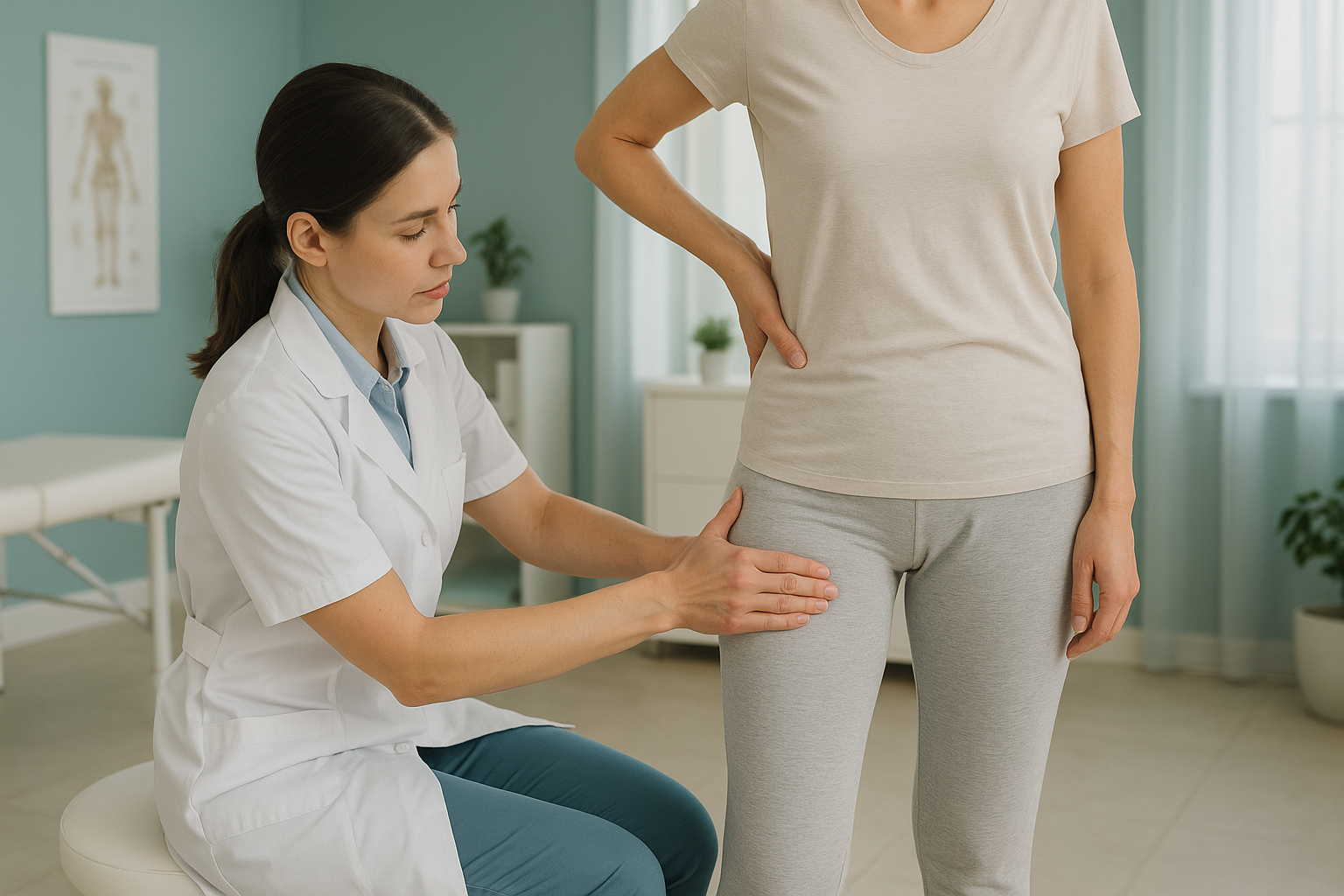
Trochanteric bursitis is pain and inflammation at the bony point on the outside of your hip, called the greater trochanter. It is often part of a broader problem now called greater trochanteric pain syndrome (GTPS), which can include gluteal tendinopathy. The pain typically worsens when you walk, climb stairs, or lie on the affected side.
Key takeaways
- GTPS is one of the most common causes of lateral hip pain in adults.
- It often involves the tendons and muscles around the hip, not just the bursa.
- Pelvic tilt, leg length difference, or altered gait can overload the outer hip.
- Standing, weight-bearing imaging can reveal alignment factors that lying-down scans miss.
- ScanAlign provides diagnostic imaging only. Treatment is guided by your clinician.
You might have been told you have trochanteric bursitis, or you might have searched "lateral hip pain" after weeks of aching that will not settle. The term is well known, but the picture is usually bigger than a single inflamed bursa. Clinicians now recognise that most outer hip pain involves changes in the gluteal tendons and surrounding structures. The umbrella term for this is greater trochanteric pain syndrome, or GTPS.
Understanding the difference matters because the right imaging can change the direction of your care. And importantly, the cause of your outer hip pain may not even start at the hip itself.
What does trochanteric bursitis feel like?
Trochanteric bursitis typically causes a deep ache or sharp tenderness on the outer side of your hip. You may also feel it radiating down the outside of your thigh. It tends to be worse at specific times.
Some people notice the pain started gradually, perhaps after increasing their walking distance or changing their exercise routine. Others cannot identify a trigger at all. Either way, the pattern of pain on the outer hip is the hallmark of GTPS.
What causes trochanteric bursitis?
GTPS is caused by excessive or repetitive load on the structures around the greater trochanter. This includes the bursae (fluid-filled cushions), the gluteal tendons, and the iliotibial band. Several factors can contribute.
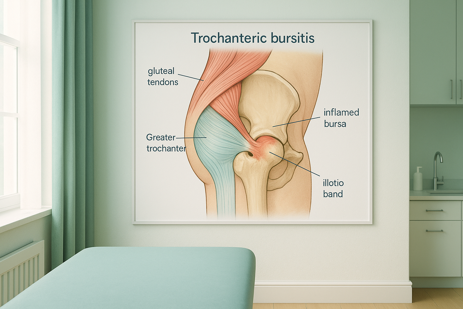
Repetitive activity such as running or hill walking is a common trigger. But in many cases, the root issue is how your body distributes load when you stand and move. A pelvic tilt, a slight difference in leg length, or a change in your walking pattern caused by a problem elsewhere can all increase strain on one trochanter. This is where whole-body alignment becomes relevant.
Why does alignment matter for lateral hip pain?
Alignment matters because your body works as a connected chain, from your feet through your knees, hips, pelvis, and spine. When one link in that chain is off, another area compensates. Pain often shows up at the point that is doing the extra work, not at the original problem.
With trochanteric bursitis, the outer hip is frequently the area absorbing extra load. A tilted pelvis may force one gluteal tendon to work harder. A stiff ankle or a knee that does not fully straighten can alter your stride and pile stress onto the greater trochanter with every step. If imaging only looks at the hip in isolation, and only while you are lying down, it may miss these contributors entirely.
Standard imaging such as X-ray, CT, or MRI is usually performed while you lie flat. In that position, gravity is removed, muscles relax, and the alignment shifts your body presents when upright and weight-bearing may disappear. That is why a scan can come back looking normal even when the pain is very real.
What helps with trochanteric bursitis?
Most treatment for trochanteric bursitis of the hip is conservative, meaning it does not involve surgery. Your clinician may recommend physiotherapy focused on strengthening the gluteal muscles, activity modification, or a temporary change in how you load the hip. Some people benefit from a steroid injection to reduce inflammation.
The key is identifying what is driving the overload. If there is an underlying alignment issue, such as a pelvic shift or a functional leg length difference, treatment can be targeted more precisely when your clinician has the full picture. That picture is what standing, weight-bearing imaging provides.
When is MRI the better choice? EOS imaging shows bones, joints, and alignment beautifully. But if your clinician suspects a soft-tissue problem such as a gluteal tendon tear, a labral issue, or nerve involvement, MRI is usually the better tool. The two types of imaging answer different questions and can complement each other.
Not sure whether your outer hip pain needs an alignment assessment? Talk to us first, at no cost.
Book a free video consultationHow does EOS imaging assess trochanteric bursitis differently?
An EOS scan captures your entire skeleton in one image while you stand naturally. This means the scan shows your real-world alignment, including how your pelvis sits, whether your legs are equal in length, and how your spine, hips, and knees relate to each other under gravity.
For someone with lateral hip pain, this can be revealing. A standard hip X-ray might show a normal-looking joint. An EOS image of the whole body might show that the pelvis is tilted, shifting extra load onto the painful side. Or it might show that a subtle knee alignment issue is changing the way you walk. These findings give your treating clinician something actionable to work with.
The EOS system also uses about 90% less radiation than a standard X-ray, around ten times less. The scan itself takes just a few seconds while you stand still in the machine. No injections, no enclosed space, no preparation needed.
ScanAlign is a diagnostic imaging clinic. We provide the imaging and the detailed report. Treatment decisions are made by your own clinician, physiotherapist, or surgeon, who can use the findings to guide your care. You can learn more about how EOS whole-body imaging works.
Common questions about trochanteric bursitis and imaging
Trochanteric bursitis specifically refers to inflammation of the bursa at the greater trochanter. GTPS (greater trochanteric pain syndrome) is the broader, more accurate term. It includes bursitis but also covers gluteal tendinopathy and other causes of lateral hip pain. Most clinicians now use GTPS because the bursa alone is rarely the whole story.
Yes. An altered gait pattern, whether from a knee problem, foot issue, or spinal condition, can change how load is distributed across your hip. Over time, this added stress on the outer hip can contribute to bursitis or tendinopathy.
It can be a contributing factor. When the pelvis is tilted, one hip may bear more load than the other. This imbalance can overwork the gluteal tendons on the loaded side and lead to pain at the greater trochanter. Standing imaging can help identify whether a pelvic tilt is present. You can read more about hip alignment assessment.
A standard hip X-ray may show bony changes near the greater trochanter, but it does not show the bursa or tendons directly. Its main role is to rule out other causes like arthritis or a fracture. An EOS standing scan adds alignment information that a standard X-ray taken lying down cannot provide.
MRI is the best tool if your clinician suspects a gluteal tendon tear or other soft-tissue damage. EOS imaging is better for assessing your skeletal alignment and how your bones and joints relate to each other when standing. The two scans answer different questions and can work together.
Yes. The EOS system uses about 90% less radiation than a standard X-ray, around ten times less. For more detail on safety, preparation, and what to expect, visit the FAQs page.
No. ScanAlign is a private, self-pay clinic. You can book directly without a GP referral. We offer a free video consultation first so we can make sure an EOS scan is the right step for you.
You can find current pricing information on the ScanAlign FAQs page.
It can contribute. Even a small difference in leg length changes the way you walk and stand. The longer leg side may take more load through the hip, increasing strain on the gluteal tendons and bursa. A full-body standing scan can measure leg length accurately under natural weight-bearing conditions.
You stand in the EOS machine wearing comfortable clothing with no metal. The scan takes a few seconds and captures your entire skeleton from the front and side simultaneously. There is no enclosed tube and no injections. Your images are reviewed by a specialist and a detailed report is produced for you and your clinician.
Wondering if your hip pain is an alignment problem?
A free video consultation lets you talk through your symptoms with our team. No commitment, no pressure.
Book a free video consultationRelated pages
Sources
- Grimaldi, A. & Fearon, A. (2015). Gluteal tendinopathy: integrating pathomechanics and clinical features in its management. Journal of Orthopaedic & Sports Physical Therapy, 45(11), 910-922.
- Segal, N.A. et al. (2007). Greater trochanteric pain syndrome: epidemiology and associated factors. Archives of Physical Medicine and Rehabilitation, 88(8), 988-992.
- NHS. Hip pain in adults. nhs.uk. Available at: https://www.nhs.uk/conditions/hip-pain/
Written by Abbas Dhami (Specialist Diagnostic Radiographer)
This page is general information and not a diagnosis.
ScanAlign is the trading name of CoreMed Solutions Ltd and operates under The Harley Street Hospital's CQC licence.