
Retrolisthesis: What Is Backward Vertebral Slippage and Should You Be Concerned?
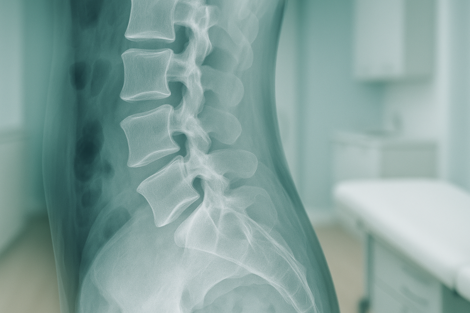
Retrolisthesis is a condition where one vertebra slips backward relative to the one below it. It most commonly affects the lower back or neck and can narrow the spaces where nerves exit the spine. Symptoms range from localised stiffness to radiating pain, and standing imaging often reveals slippage that lying-down scans underestimate.
Key takeaways
- Retrolisthesis means a vertebra has shifted backward out of its normal position.
- It is graded by how far the bone has moved, from mild (grade 1) to more significant.
- Lying-down scans can hide the true extent of slippage because gravity and body weight are removed.
- Standing, weight-bearing imaging shows your spine as it functions in daily life.
- If you develop numbness, weakness in your legs, or loss of bladder or bowel control, seek urgent medical attention (GP, 111, or A&E).
Your spine is made up of individual bones (vertebrae) stacked on top of each other. Normally they line up neatly in a gentle curve. With retrolisthesis, one of those bones slides backward, even by a small amount. That shift can narrow the channel your spinal cord runs through and squeeze the openings where nerves branch out to the rest of your body.
The condition is often found on an X-ray or scan taken for back or neck pain. You might see terms like "grade 1 retrolisthesis" on a report, which simply means the slippage is at the milder end of the scale. Higher grades indicate more movement. The grading helps clinicians decide how best to manage it.
What are the symptoms of retrolisthesis?
The most common symptom is a dull, aching pain in the area of the slippage, often in the lower back or neck. Some people also feel stiffness, restricted movement, or pain that travels into the buttocks, legs, or arms depending on which part of the spine is affected.
Not everyone with retrolisthesis has symptoms. Some people only discover it when they are scanned for another reason. The severity of symptoms does not always match the grade of slip, which is why a full picture of your alignment matters.
What causes retrolisthesis?
Retrolisthesis develops when the structures that hold a vertebra in place lose strength or become damaged. The most common cause is disc degeneration, where the cushion between two vertebrae thins over time, allowing the bone above to slide backward.
Age-related changes are the most frequent contributor, but retrolisthesis can also follow surgery at a nearby spinal level or develop alongside other conditions like scoliosis. Understanding the root cause helps shape how it is managed.
What is the difference between retrolisthesis and spondylolisthesis?
The key difference is direction. In retrolisthesis the vertebra slips backward, while in spondylolisthesis it slips forward. Both involve one bone moving out of line with its neighbour, and both can compress nerves. Spondylolisthesis is more widely discussed, but retrolisthesis is not rare and can cause similar symptoms.
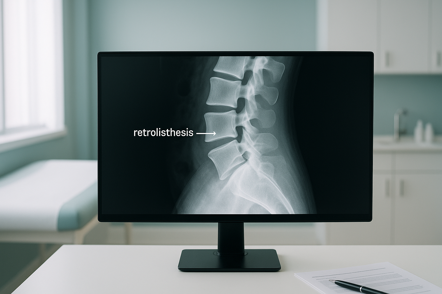
Both conditions are best assessed with the spine upright and under load. When you lie down for a standard scan, gravity is removed and muscles relax. A slip that causes problems while you are standing or walking may look less significant, or even disappear, on a lying-down image.
How does standing imaging help assess retrolisthesis?
Standing, weight-bearing imaging captures your spine exactly as it works in everyday life, with gravity pulling on it and your muscles actively supporting it. This reveals the true position of each vertebra under load, which is critical for a condition defined by movement between bones.
EOS is a low-dose imaging system that scans your entire body while you stand. It uses about 90% less radiation than a standard X-ray, around ten times less. Because it captures your full skeleton from head to feet in one image, your clinician can see how a slip at one spinal level relates to the alignment of your pelvis, hips, and legs below.
Your body is a connected chain. A backward slip in the lower back changes the curve of your spine, which can tilt your pelvis, shift your weight, and create strain in your hips or knees. Conversely, a problem lower down, like a leg-length difference, can contribute to retrolisthesis higher up. Whole-body standing imaging shows these connections in a way that single-area, lying-down scans simply cannot.
Want to understand how your spine alignment looks under real-world conditions?
Book a free video consultationWhat can be done about retrolisthesis?
Management depends on the severity and symptoms. Mild cases are often managed conservatively with physiotherapy to strengthen the muscles that stabilise the spine, particularly the deep core and back extensors. Activity modification and postural awareness also play an important role.
More significant or symptomatic cases may need input from a spinal specialist, who might recommend targeted injections or, in some situations, surgery. The first step in any pathway is an accurate picture of what is happening. Knowing the grade and direction of slip, whether neighbouring levels are affected, and how the rest of the skeleton has adapted all help your clinician choose the right approach.
When is MRI more suitable? EOS excels at showing bone position, joint alignment, and the overall balance of your skeleton. If your clinician suspects disc herniation, nerve compression, or other soft-tissue problems alongside retrolisthesis, MRI is usually the better tool for those specific questions. The two scans complement each other well.
Frequently asked questions about retrolisthesis
It depends on the grade and whether it is pressing on nerves. Many people have mild retrolisthesis with few or no symptoms. If you develop numbness, weakness in your legs, or loss of bladder or bowel control, seek urgent medical attention (GP, 111, or A&E).
Grade 1 means the vertebra has slipped backward by a relatively small amount. It is the mildest grade and is often found incidentally on imaging. Many people with grade 1 retrolisthesis manage it successfully with strengthening exercises and monitoring.
It can, particularly if the underlying cause (such as disc degeneration or joint wear) progresses. Regular assessment helps track any change, and strengthening the muscles around the spine can slow progression.
Core stabilisation, pelvic tilts, and controlled extension exercises are commonly recommended. A physiotherapist can design a programme specific to your level of slip and symptoms. Accurate imaging beforehand helps them target the right area.
Retrolisthesis is about the position of the bone, while a herniated disc is about the soft cushion between the bones bulging or rupturing. They can occur together, and both can compress nerves, but they require different types of imaging to assess properly.
When you lie flat, gravity no longer pulls on your spine and your muscles relax. The slipped vertebra can settle back closer to its normal position, making the slip look smaller or even invisible. Standing imaging shows the slip under real-world load.
Yes. EOS uses about 90% less radiation than a standard X-ray, around ten times less. You can find more details on safety and what to expect on our FAQs page.
No. ScanAlign accepts self-referrals. You can book a free video consultation directly and discuss whether an EOS scan is appropriate for you.
Pricing details are available on our FAQs and cost page.
Yes. A backward slip changes how your spine curves, which can alter your pelvic tilt and shift weight unevenly through your hips and knees. This is why whole-body imaging is valuable. You can learn more about how hip alignment connects to the spine.
Understand your spine under real-world conditions
A free video consultation lets you discuss your symptoms and find out whether standing, whole-body imaging is right for you. No referral needed.
Book a free video consultationWritten by Abbas Dhami (Specialist Diagnostic Radiographer)
Sources
- NHS. "Back pain." nhs.uk, accessed 2024.
- Shen M, Razi A, Engquist D. "Retrolisthesis and lumbar disc herniation: a preoperative assessment of patient function." The Spine Journal, 2007;7(4):406–413.
- American Academy of Orthopaedic Surgeons (AAOS). "Spondylolisthesis." orthoinfo.aaos.org, accessed 2024.
Learn more about how standing spine imaging reveals alignment problems, or explore the full EOS scan overview.
This page is general information and not a diagnosis.
ScanAlign is the trading name of CoreMed Solutions Ltd and operates under The Harley Street Hospital's CQC licence.